Slika:Gray491.png
Gray491.png (500 × 438 točk, velikost datoteke: 63 KB, MIME-vrsta: image/png)
Spodaj prikazane informacije so s tamkajšnje opisne strani.
Povzetek
| Opis |
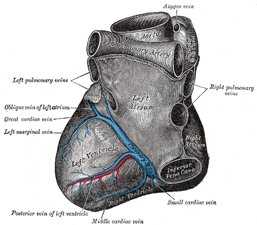
Deutsch: Blick von hinten auf das Herz. Darstellung von Henry Gray. |
||||||||||||||||||||
| Plošča | 491 | ||||||||||||||||||||
| Datum | pred letom 1858 | ||||||||||||||||||||
| Vir |
|
||||||||||||||||||||
| Avtor |
|
||||||||||||||||||||
.jpg)
Knjiga
| Henry Gray: Gray's Anatomy (20. izdaja)
|
|||||||||||||||||||||||
|---|---|---|---|---|---|---|---|---|---|---|---|---|---|---|---|---|---|---|---|---|---|---|---|
| Avtor |
|
-_Title_page.png) | |||||||||||||||||||||
| Urednik |
Revised by Warren H. Lewis |
||||||||||||||||||||||
| Ilustrator |
|
||||||||||||||||||||||
| Naslov | |||||||||||||||||||||||
| Izdaja |
20 |
||||||||||||||||||||||
| Izdajatelj | |||||||||||||||||||||||
| Vrsta objekta |
izdaja |
||||||||||||||||||||||
| Pregled strani | list of all the plates | ||||||||||||||||||||||
| Jezik |
angleščina |
||||||||||||||||||||||
| Datum objave |
1918 |
||||||||||||||||||||||
| Kraj izida |
Filadelfija / New York |
||||||||||||||||||||||
| Vir | Bartleby | ||||||||||||||||||||||

Licenca
Slika je v javni domeni, saj je zgolj mehanska preslikava ali fotokopija izvirnika, ki je v javni domeni, ali pa je - po dostopnih informacijah - preveč podobna taki preslikavi ali fotokopiji, da bi bila lahko zaščitena z avtorskimi pravicami. Izvirnik sam je v javni domeni iz naslednjega razloga:
Oznaka je prirejena za uporabo v primerih, ko je treba izjaviti, da je vsaka izboljšava (npr. poprava osvetlitve, kontrasta, barv, ostrine) sama po sebi nezadostna, da bi tvorila nove avtorske pravice. Uporabi se lahko povsod, kjer ni znano, ali so bile napravljene izboljšave, kot tudi takrat, ko so izboljšave jasne, vendar ne zadostujejo za novo avtorsko delo. Za znane neobdelane preslikave lahko uporabite oznako {{PD-old}}. Uporaba predloge {{PD-scan}} je opisana na strani Commons:When to use the PD-scan tag.  | ||||
The coronary sinus is a collection of veins joined together to form a large vessel that collects blood from the myocardium of the heart. It is present in humans and other animals. It delivers deoxygenated blood to the Right atrium in conjunction with the superior and inferior vena cava.
The coronary sinus opens into the right atrium, between the inferior vena cava and the atrio-ventricular orifice. It returns the blood from the substance of the heart, and is protected by a semicircular fold of the lining membrane of the auricle, the coronary valve (the valve of Thebesius). The sinus, before entering the auricle, is considerably dilated - nearly to the size of the end of the little finger. Its wall is partly muscular, and at its junction with the great coronary vein is somewhat constricted and furnished with a valve consisting of two unequal segments.(Gray 462)
Location: It is located in the right atrium and runs transversely in the groove between the left atrium and ventricle on the posterior surface of the heart.
The coronary sinus orifice (opening) is just superior to the septal leaflet of the tricuspid valve. The coronary sinus orifice is also known as the ostium of the coronary sinus, and is guarded by the Thebesian valve.
Drainage: It receives blood mainly from the small, middle, great and oblique cardiac veins. It also receives blood from the left marginal vein and the left posterior ventricular vein. The anterior cardiac veins drain directly into the right atrium. (Some small veins drain into any of the four chambers of the heart.)
It drains into the right atrium on the posterior, inferior surface, medial to the inferior vena cava opening.
Zgodovina datoteke
Kliknite datum in čas za ogled datoteke, ki je bila takrat naložena.
| Datum in čas | Sličica | Velikost | Uporabnik | Komentar | |
|---|---|---|---|---|---|
| trenutno | 22:35, 23. januar 2007 | | 500 × 438 (63 KB) | Pngbot | optimized with optipng |
| 08:26, 11. februar 2006 |  | 500 × 438 (100 KB) | Arcadian | {{Gray's Anatomy plate}} |
Uporaba datoteke
Datoteka je del naslednje 1 strani slovenske Wikipedije (strani drugih projektov niso navedene):
Globalna uporaba datoteke
To datoteko uporabljajo tudi naslednji vikiji:
- Uporaba na ar.wikipedia.org
- Uporaba na bg.wikipedia.org
- Uporaba na bn.wikipedia.org
- Uporaba na bs.wikipedia.org
- Uporaba na cv.wikipedia.org
- Uporaba na de.wikibooks.org
- Uporaba na el.wikipedia.org
- Uporaba na en.wikipedia.org
- Coronary circulation
- Coronary sinus
- Oblique vein of the left atrium
- Posterior descending artery
- Circumflex branch of left coronary artery
- Vital heat
- Posterior interventricular sulcus
- Left marginal artery
- Smallest cardiac veins
- Vascular remodelling in the embryo
- Crux cordis
- User:Bob K31416/BH
- User:Walkerc84/sandbox
- User:Was a bee/Gray
- Uporaba na es.wikipedia.org
- Uporaba na fa.wikipedia.org
- Uporaba na it.wikipedia.org
- Uporaba na ja.wikipedia.org
- Uporaba na ko.wikipedia.org
- Uporaba na nl.wikipedia.org
- Uporaba na nn.wikipedia.org
- Uporaba na pl.wikipedia.org
Oglejte si globalno uporabo te datoteke.